2020 Imaging Tethered DR Panels
.
P-DR 1012V 10x12in Tethered Flat Panel DR Detector
PRODUCT CODE: P-DR-1012V

Taking digital radiography to the next level with our tethered Flat panel technology is 2020 Imaging’s cassette-sized 1012V. Providing exceptional image quality at 16-bit, paired with advanced software features and functionality. The 10×12 panel uses enhanced Cesium technology and has many unique features to fit the needs of your office configuration.
- Warranty: 3 years
- Imaging Area: 10″ x 12″
- Line pair per mm: 4.0
- Cassette sized
- 5.5 lbs
- Pixel Matrix: 2000×2400
- Off-center Imaging
- A/D conversion: 16-bit
Multimedia:
.
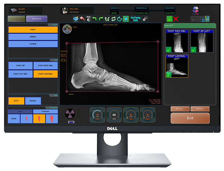
2020 Imaging Acquisition Workstation
PRODUCT CODE: 2020I-AWS
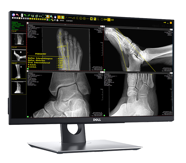
State-of-the-art DICOM viewer delivers powerful, intuitive workstation functionality
Quick function shortcuts integrated for efficiency, Podiatric (DPM) Tools available
WORLDWIDE access: view from anywhere!, High-resolution multi-monitor support, 4k monitor support
Customize screen layouts; up to 9 images per monitor. Fully customizable settings to accommodate your specific needs, Compare images (post/pre op), Refresh (see saved/available images, while study is being performed), Custom Toolbox (see top-left of above image), fully customizable annotations/tools
Bone Enhancement – reprocess images sharper for enhanced diagnosis
Software Integration
- Acquire Software/Generator Integration.
- Anatomical Program Selection Preset x-ray techniques & exams.
- Selection directly at workstation.
- Superior Image Quality, Sharper images, optimized clarity.
- Quick view selection.
- Customizable image sharpness level.
- Auto Contrast upon processing, Image auto-shutter/crop.
- Images auto-save during acquisition to Server for immediate access in exam rooms.
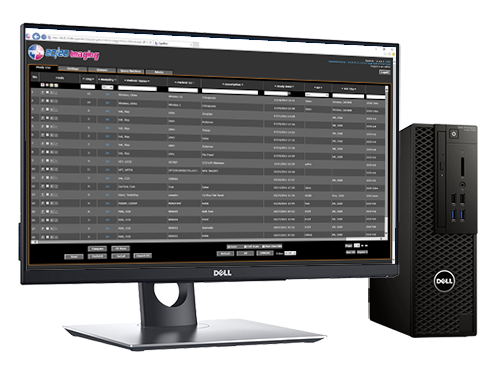
- Business Class Components, Dell Server PC (Full PACS)*, Core i5, 8GB ram
- 1TB RAID 1 mirror (failsafe system)
- Space saving Small Form Factor design
- Dell 24″ LED IPS 2MP Touchscreen Monitor, VESA Wall-mount compatible
- 10-point Touchscreen, High Quality Display for optimized viewing
Opal-RAD Software
- Multi-functional Studylist (see above), Burn/Import CDs
- Compare images (post/pre op), Custom search/query
- Customize user privileges, Acquisition (see above)
- Processing Technology: sharper images, clarity
- Auto Contrast upon processing, Image auto-shutter/crop
- User friendly, DICOM compliant, DPM Tools
- 5 additional viewing licenses included
- WORLDWIDE access: view from anywhere with internet access.
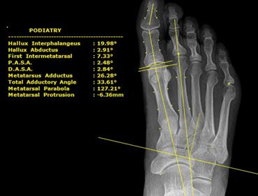
Podiatric Tool-Set
- Hallux Interphalangeus, Hallux Abductus, First Intermetatarsal, P.A.S.A., D.A.S.A., Metatarsus Adductus, Total Adductory Angle, Metatarsal Parabola, Metatarsal Protrusion, Talo-Calcaneal
Realtime Cloud Backup
- Add Cloud-Backup at any time! Add’l fee applies***
- Live synchronization in the background
- Secure 448-bit encryption for all data traffic
- Enterprise grade software (CrashPlan Enterprise)
- Customizable to meet office bandwidth needs
- Safe & Secure, Worry-Free
mOpal Software:
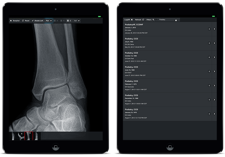
IPAD®, IPHONE®, ANDROID™, BLACKBERRY®, ANY MOBILE OR SMART DEVICE!
Combine the versatile Opal-RAD Professional Workstation software with our web-based DICOM Viewer and you have Anywhere, Anytime Viewing now on Any Device.
mOpal* (mobile Opal) is a browser-based viewer for reviewing images on Mac, Windows, mobile devices, tablets and smart phones including iPad, iPhone, Android, and Blackberry. With mOpal, receive fast access both onsite or remotely**, available with any 20/20 system.
- Can be accessed on tablets, Macs, and mobile phones
- Perfect for exam room viewing or offsite consults
- Zero footprint, no download or app needed
- Great for referring physicians
- Same user-name and password as 20/20 Imaging PACS
- Patient list and images available for anytime viewing
- Simple patient filter and search
- View annotations created on OpalWeb
- Fully customizable settings to accommodate your specific needs
- Easy to use: tap, drag, and pinch movements to manipulate images
Multimedia:
.


