Del Medical Apollo DRF Advanced Remote Controlled R/F System
PRODUCT CODE: APOLLO-DRF
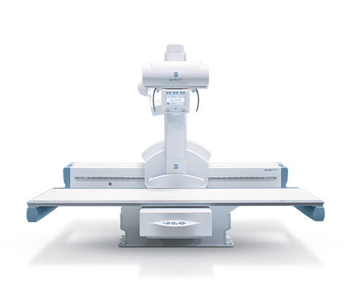
Apollo DRF is Villa’s reference product in the panorama of digital remote-controlled tables. The Apollo DRF stands out over time for both its unique, and equally innovative features. Its top performance and operability have always been widely acknowledged. With features such as wide application flexibility, high productivity and high image quality, the Apollo DRF is still one of the most appreciated products used by radiographic professionals today.
This new version of the Apollo DRF remote-controlled table has been equipped with a number of innovative features that significantly improve its performance and make its use more straightforward and user-friendly. This maximizes the efficiency of daily routine usage.
Implementing new examination procedures in the system also allows additional diagnostic information to be acquired, thus making the diagnosis process more comprehensive. With this new version, Apollo DRF achieves extraordinary application flexibility. Combined with high performance the Apollo DRF redefines the users experience of the system.
Superlative Flexibility
All Apollo DRF movements have been designed to ensure rapid and accurate positioning and allow the widest possible range of radiographic projections. The 90° tilt in both directions makes it possible to install the remote-controlled table in the most diverse configurations of the diagnostic room. This allows top results to be obtained in any environment. The wide travel of the tube-detector assembly ensures complete patient coverage, thus avoiding the need for its repositioning or to move the table longitudinally.
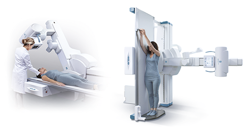
By means of motorized tilting of the tube support column, combined with rotation of the X-ray source, oblique projections can also be carried out on the table as well as patient exposures on a stretcher. The focus-detector distance, which can be extended up to 180 cm, makes it possible to examine the chest directly on the remote-controlled table.
The lower height from the floor and the completely smooth surface of the examination table make patient access easier and simplify transfer procedures from the stretcher. In addition, the sturdy and reliable mechanical structure can withstand a weight up to 284 kg with no movement restrictions, thus also making it possible to examine bariatric patients.
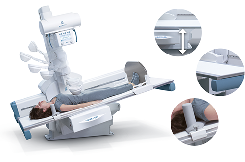
The system can be supplied with a wide range of accessories for patient positioning and for special procedures, such as lateral cassette holder, compression band, shoulder support, patient footrest, handles and leg support.
Efficiency with a Simple Touch
Thanks to the seamless integration of the remote-controlled table with the digital acquisition system, Apollo DRF simplifies and speeds up the workflow, reducing the need for manual operations by the operator. In fact, the system recognizes the type of projection to be performed and automatically adjusts all examination parameters according to the pre-set values for each projection. By simply pressing a key the table automatically reaches the preset position setting all geometric features according to the radiographic projection to be performed, such as focus-detector distance, collimation area, additional filtration and correct anti-scatter grid between the two available in the system.
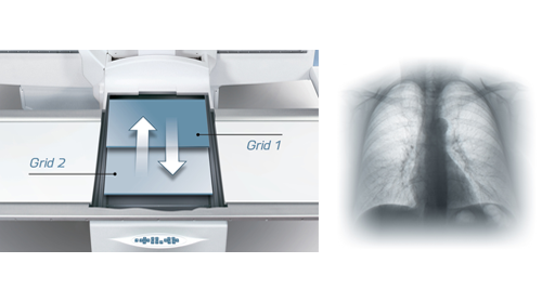
Should grid use not be required, such as when examining pediatric patients or extremities, the system automatically parks the grid outside of the X-ray field, thus resulting in a reduction of the dose delivered.
The system provides the operator with significant flexibility in controlling movements of the table through the various interfaces available. The remote-control console, which is based on an upgraded high performance hardware architecture, enables user-friendly control of the equipment via the large touch screen display that instantly shows information regarding table position and operating mode.
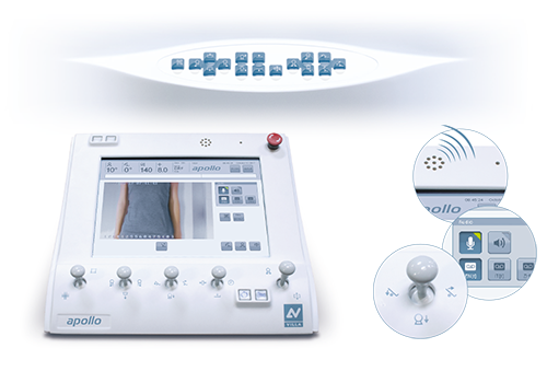
The smart-touch joysticks, which are activated by human touch, control all motorized movements in total safety, thus adding a further prevention feature against accidental activation. The operator is also able to communicate directly with the patient to provide instructions and reassurance during the exam via the built-in two-way intercom, which is supported by pre-recordable audio messages in several languages. The video camera built into the collimator makes it possible to perform the centering operation without X-ray emission and provides a new visual of the patient during positioning.
Should it be required to stay close to the patient when preparing the examination, remote controlled table movements can be driven from the control panel on the side of the patient tabletop or via an additional control panel on a trolley.
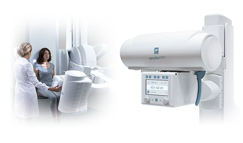
The operator can also perform centering operations directly from the new collimator with the touch screen interface, from which the position of the X-ray source on the anatomical part concerned can be adjusted. The collimator display also shows information regarding SID, the collimation area and additional filtration. Apollo DRF therefore provides the user with high-level ergonomics, resulting in rapid and efficient workflows.
High-quality Images and High Productivity
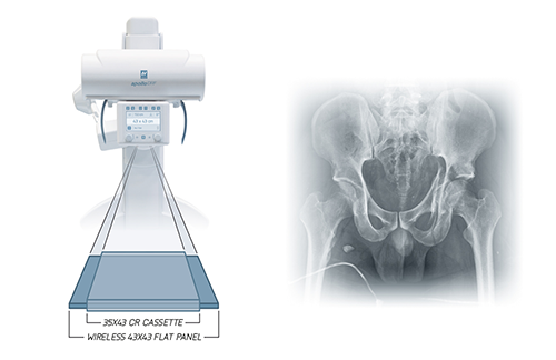
The large dynamic detector provides high-resolution X-ray images and high-acquisition frequency fluoroscopic sequences, rapidly switching between modes. Its high resolution and sensitivity allow the system to obtain extremely detailed and sharp images at low dose, while the active area of 43×43 cm assures the right coverage of each anatomical area.
All that makes Apollo DRF a “2-in-1” solution able to cover a wide range of diagnostic applications with extreme versatility, constantly maintaining high quality levels.
The digital acquisition system features a new user graphic interface*, which is entirely redesigned for quick and user-friendly use: the screen has in fact been divided into designated areas for setting examination parameters, acquired image display and application of post-processing tools.
Moreover, the image processing algorithm has been upgraded, making it even more accurate in automatically identifying the processing parameters of the anatomical area examined to optimize visibility of the details. High diagnostic content images are thus obtained just a few seconds after exposure, making the diagnostic process even safer and more effective.
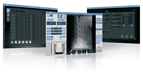
*available from second quarter 2018
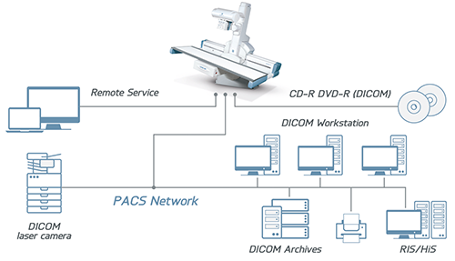
As a result of the fully available DICOM functions, the Apollo DRF’s acquisition system efficiently interfaces with the HIS/RIS and PACS structures, assuring the best use of digital technology’s full potential.
An example of integration between the various application environments is given by the auto-positioning function that Apollo DRF is equipped with: by automatic patient/examination list reception, the remote controlled table’s software is able to set up the generator’s exposure parameters, position the remote controlled table’s geometry according to protocol and make the images available to diagnostic workstations immediately after exposure. This RIS mapping function boosts the workflow and results in a considerable increase in production.
A Wide Range of Applications
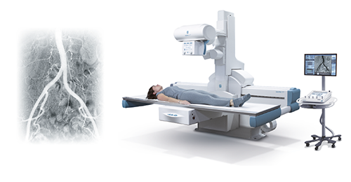
The significant versatility of Apollo DRF is expressed in its ability to cover a wide range of routine and specialized examinations, streamlining use and productivity of the R/F examination room. The large active area of the detector and high quality of the acquired images are conducive to an extraordinary range of possible applications in radiography and fluoroscopy: from general radiology procedures to dynamic ones focusing on the digestive or peripheral venous system, from pain therapy procedures to micro-invasive procedures, from urography to tomography examinations. In addition, vascular examinations are also possible via the digital subtraction angiography (DSA) package.
Advanced Functions for Effective Diagnoses
In the configuration with an additional wireless detector which may be seamlessly integrated with the same digital acquisition system of the remote-controlled table, the application abilities of Apollo DRF are further extended making it possible to perform exposures in direct contact with the detector and on stretcher patients.
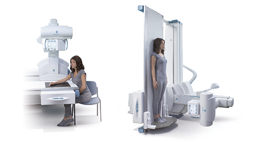
This operational flexibility may be further extended by using a second X-ray tube on a suspended ceiling stand that can be controlled by the same remote-control system generator which, together with the wireless detector, also makes it possible to perform lateral projections on the table.
Furthermore, the stitching procedure, which consists in automatic acquisition process of a set of X-ray exposures and their subsequent integration into a single image, is also available. This function therefore supports investigation of extensive body areas, such as full-leg/full-spine examinations, also extending diagnostic capabilities to orthopedics thanks to the optional specialist measurement package.
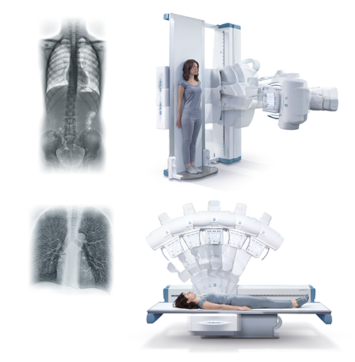
In addition, Apollo DRF now features the innovative tomosynthesis function, an examination technique based on low-dose acquisition of a series of projections from different angles of an anatomical part, which are processed through advanced reconstruction algorithms to obtain a sequence of slices parallel to the examination table. The images obtained, free from overlap by the surrounding tissues, allow one to more easily determine the presence of small lesions or anomalies that are not very visible with conventional radiography, thus providing information to make the diagnostic process faster and more effective.
Evolution that Makes the Difference
- Apollo DRF features a number of solutions that are thoroughly designed to reduce the dose and to assure the utmost safety for the patient:
- New multi-layer table surface with reduced X-ray attenuation
- Automatic removal of the anti-scatter grid for examinations of pediatric patients and extremities
- Automatic collimator available with optional additional filters
- Anatomic programs optimized for the body part to be examined, with specific mode for pediatric patients
- Dynamic Flat Panel Detector with high X-ray sensitivity
- Pulsed-fluoro mode with variable acquisition speed
- Virtual collimator function with diaphragm adjustment without X-ray emission by using the last image hold (LIH)
- Virtual scan function that displays movement of the collimator on the last image acquired during table movements, thus making it possible to center the area of interest without X-ray emission
- Other features assure additional safety for the patient as well as the operator during the examination:
- Smart-touch joysticks, equipped with a proximity sensor that reduces involuntary activation, to control the motorized movements in utter safety
- Software designed to monitor all movements of the equipment, supporting the utmost operator confidence
- High-voltage cables encased inside the table’s covers so that examinations may be carried out with peace of mind.

