
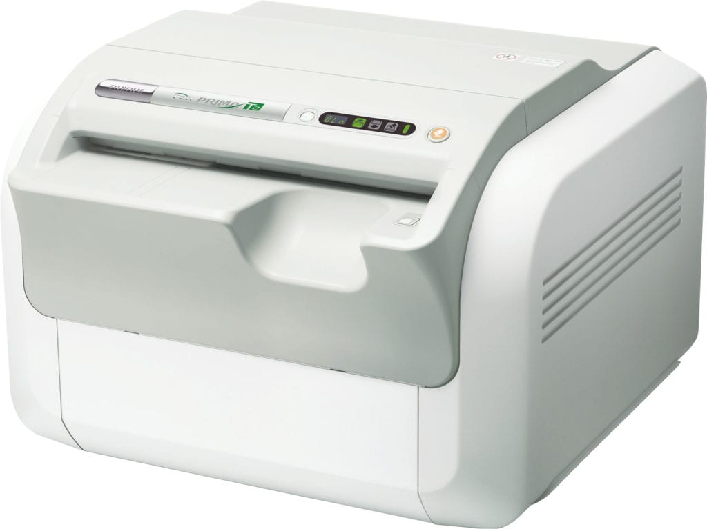
FUJIFILM FCR Prima T2
PRODUCT CODE: FCRPRIMAT2
Ideal for Private Practices
FCR Prima T2 has been designed for those private practitioners with low volume imaging environments. Small, light, fast, it has everything a smart private practice wants in digital x-ray. At just 86 pounds and with a footprint of only 3.2 square feet, it’s one of the smallest table top CR systems available.
Impressive Speed and Image Quality
The FCR Prima T2 stands out for its stunning combination of price, speed and remarkable image quality. Its throughput ranges from a rapid 47 to an outstanding 73 images per hour depending on cassette size. Its 100-micron images provide impressive resolution while Fujifilm’s processing technology ensures your first image is ideal.
Workstation Flexibility
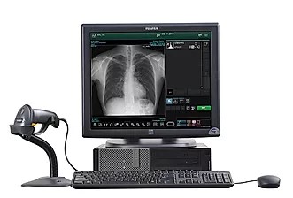
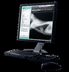
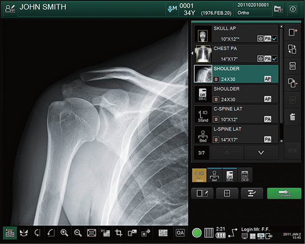
FCR Prima T2 is available with the FDX Console and FCRView workstations. The FDX Console is Fujifilm’s most cutting edge acquisition workstation, designed to streamline the operation of digital image acquisition products. It features Fujifilm’s most advanced image processing: Dynamic Visualization™. The FCRView is a complete image management solution that provides users with a comprehensive set of tools to acquire,view and archive images all from one workstation.
Technical Specifications:
FCR Prima T2
IP Cassette Type III-CC
- – 14 x 17″ (35 x 45 cm)
- – 14 x 14″ (35 x 35 cm)
- – 10 x 12″ (24 x 30 cm)
- – 8 x 10″
- – 15 x 30 cm
Time required for IP feed/ load: Min. 49 sec. (14 x 17″)
Processing capacity: Up to 73 IPs/hr
Reading specification: 10 pixels/ mm, 5 pixels/ mm
Time to start on display: Min. 33 sec.
Footprint: Less than 2.5 sq. ft.
Width: 22″ (56 cm)
Depth: 21″ (54 cm)
Height: 15″ (39.2 cm)
Weight: 86 lbs. (39 kg)
Power requirements: Single phase 50- 60 Hz, AC 120-240 V ±10% 1.9A (max)
Multimedia:
.
FDX CONSOLE
PRODUCT CODE: FDXCONSOLE
The FDX Console is the heart of every FDR and FCR system. It represents the culmination of Fujifilm’s extensive experience in image and information processing. The sophisticated interface is intuitive and customizable, and enables fast, easy exam completion and image optimization
New features include:
- Dynamic Visualization™ is Fujifilm’s advanced image processing technology, providing outstanding first-up detail and virtually eliminating the need for post processing.
- Complete common workflow steps in as few as 2-3 mouse clicks
- Worklist views with status icons and thumbnail images
- Virtual Grid™ (option) – improved image quality for images acquired without a grid
- Double click full-screen zoom
- PICC line/edge enhancement toggle
- Dose tracking and management tools
- Optional 2MP secondary monitor for PACS comparable preview
Dose optimization features
- Customizable menus help simplify dose-saving techniques.
- Exposure Index (EI) and Deviation Index (DI) guides and tracks deviation from preferred exposure conditions for each exam.
- EI and DI values can be sent in DICOM headers, allowing low dose initiative monitoring.
See more, click less
- Multi-Accession opens multiple studies with separate accessions for the same patient in one acquisition screen
- Auto trimming simplifies offset imaging by automatically detecting the exposure area. The true sized cropped image is sent to PACS, for optimal image display size at both workstations.
- Region of Interest (ROI) image adjustment — unique 2-point ROI function that reprocesses images instantly based on user-selected points of interest.
- Image Stitching (optional) for DR and CR automatically combines multiple adjoining images into a single long length image for scoliosis and long leg studies.
Accessory
Wall mounting hardware for FDX Console
Delivering a refined image processing experience
Dynamic Visualization™ processing tools produce high quality images that aid diagnosis and boost productivity. Optimal first-up display virtually eliminates any need for post processing image adjustments, providing exceptional image quality automatically. Wide dynamic range adaptability and breadth of exam menus help to reduce retakes.
Fujifilm’s Dynamic Visualization image processing automatically recognizes the region of interest and applies the optimum image processing parameters throughout the entire exposure field producing exceptional images with higher window and leveling content for faster, more accurate diagnosis. Additional advanced functions include:
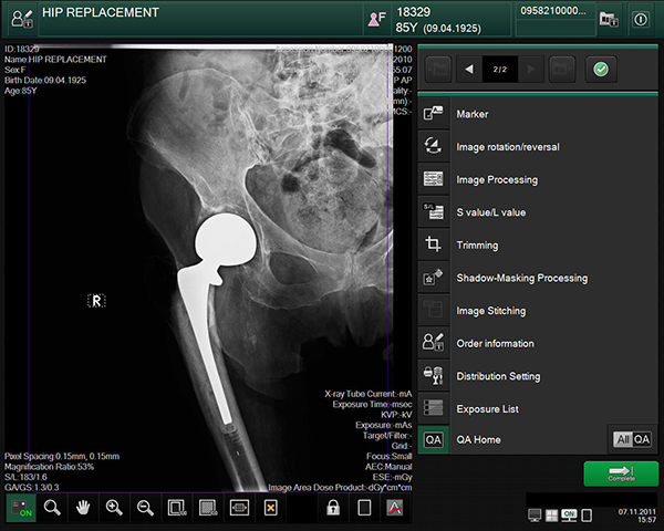
- Dynamic Visualization™ II (upgrade option): Our latest evolution in image processing. Dynamic Visualization II advanced processing with auto recognition of bone, anatomy characteristics, and orthopedic hardware. This image processing software intelligently adapts image contrast and density, based on image, thickness and structural recognition. It improves uniformity in both dense and thin regions for challenging images, for large anatomy and patients, or any low dose or low penetration exams.
- Virtual Grid (option): Processing intelligently simulates grid use, eliminating scatter effect, to improve contrast and clarity for images acquired without a grid. Useful in portable exams: simplifies acquisition and positioning, and eliminates artifacts associated with physical grid misalignment and improper SID.
- Intuitively recognizes scatter effect in the image
- Precisely tunes contrast and noise control
- Eliminates misalignment factors
- Customizable grid characteristics, grid lines, density and interspacing material
- Multi-Frequency Processing (MFP): Applies edge enhancement and gray scale processing to multiple frequencies, improving visibility for varying densities and foreign structures. Useful in viewing spine, skull and orthopedic hardware images
- Flexible Noise Control (FNC): Selectively suppresses noise without loss of sharpness. Useful for pediatrics, lumbar spine and abdomen views
- Grid Pattern Removal (GPR): Intelligently removes moiré patterns caused by grids
- Fujifilm DR to Fujifilm Synapse PACS viewing shortcuts: Applies radiologist-specific single-click processing preferences to views, significantly simplifying workflow
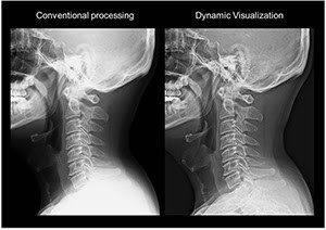
Conventional Processing vs Dynamic Visualization 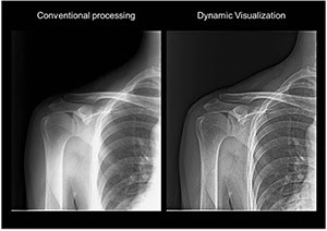
Conventional Processing vs Dynamic Visualization 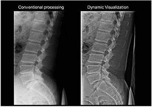
Conventional Processing vs Dynamic Visualization 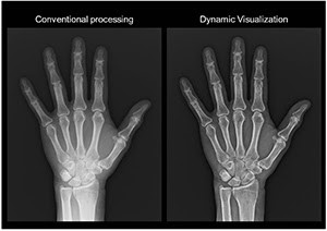
Conventional Processing vs Dynamic Visualization 
Conventional Processing vs Dynamic Visualization 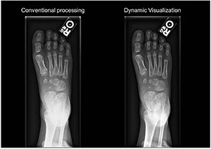
Conventional Processing vs Dynamic Visualization 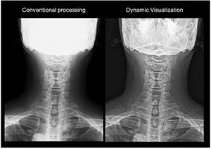
Conventional Processing vs Dynamic Visualization

Virtual Grid™
As the leader in digital radiography image processing, Fujifilm is proud to introduce Virtual Grid. Virtual Grid processing corrects for the effects of scatter radiation that would otherwise reduce image contrast and clarity for images acquired without an anti-scatter grid.
Intelligent image processing that replaces the use of a grid
Virtual Grid™ processing enhances image contrast and clarity with up to 50% dose reduction compared to a real grid. Physical grids are commonly required for mobile imaging of large anatomy to help focus radiation and reduce scatter. Virtual Grid processing will be of great benefit to technologists for mobile imaging applications in emergency room, operating room, critical care and other exams. Virtual Grid can be applied to all body parts,* including chest, abdomen, head, spine, pelvis, upper and lower extremities.
With the ability to customize its emulated grid characteristics, Virtual Grid provides exam flexibility and eliminates image quality problems that result from improper grid alignment or focus.
By simulating actual grid use, Virtual Grid can be beneficial in many clinical scenarios (bedside, ER, OR, ICU) where positioning a physical grid can be challenging or disruptive to patient comfort.
- Eliminates physical grid-related misalignment issues
- Precisely tunes image contrast while suppressing image noise
- Emulates a wide range of physical grid characteristics, by grid ratio, density and interspace material
*Excluding breast imaging.
Virtual Grid requires FDX Console Version 9 or greater application software.
Scoliosis and Image Stitching
Fujifilm’s 10″ x 24″, 14″ x 34″ and 14″ x 50″ Long View Cassettes allow the imaging plates (IPs) to be loaded and processed in the FCR reader, without manually removing them from the cassette. The lightweight and simplified design makes it easy for technologists to perform specialized applications without interruptions. All cassettes use a patented design featuring latches at either end of the cassette to promote ease of workflow. Imaging plates (IPs) inside the cassettes overlap by 1″ to ensure that no pathology is lost. The cassettes include markers which allow Fuji’s stitching software to automatically compose images. Grids, wall stands and IPs are available separately from the cassette.
- 10″ x 24″ cassette consists of two interlocked 10″ x 12″ cassettes and requires two 10″ x 12″ IPs for use. Ideal for smaller patients.
- 14″ x 34″ cassette consists of two interlocked 14″ x 17″ cassettes and requires two 14″ x 17″ IPs for use. Excellent for most scoliosis applications.
- 14″ x 50″ cassette consists of a special 14″ x 17″ cassette which can be inserted into a 14″ x 34″ cassette and attached using a simple latch on either side for easy removal for processing. Three 14″ x 17″ IPs are required for this cassette. Ideal for long leg studies or other applications where a wider view is required.
Fujifilm Automatic Image Stitching Software
Fujifilm’s Automatic Image Stitching Software enables display of the entire spine or lower extremities on a single image.
- Composes up to three individual images.
- Automatically mergers images together by aligning markers on the image. Manual adjustments can be applied if necessary.
- Image processing is applied and optimized to the resulting image display.
- Simply select the proper menu and the Flash IIP console Autostitch software can take care of the rest.
Advanced Image Processing
Issues often associated with image stitching software include difficulty with image alignment and variations of density on the composed image. Fujifilm’s comprehensive, advanced image composition processing eliminates manual alignment requirements and compensates for differences in density in the image. Fujifilm’s Image Stitching Software enables faster exam processing, higher diagnostic confidence and quicker image interpretation.
Fujifilm Type CC Cassettes
Fujifilm CR cassettes are available in the most common sizes as well as extended lengths for long leg and scoliosis exams. The cassettes are of a lightweight and rugged design to safely house and protect Fuji’s advanced flexible imaging plates. Depending upon the package, Cassettes and IPs are included and also are sold separately.
Fujifilm Imaging Plates (IPs)
Fujifilm Imaging Plates (IPs) are the result of years of research and continuous improvement. Fujifilm’s IP design employs an extremely efficient image recording medium based on a high-emission, photo-stimuable phosphor, which accurately detects and stores x-ray energy in its phosphor particles. Fujifilm’s phosphor composition offers a high sensitivity, high sharpness and low noise. Its remarkable luminescence linearity and broad sensitivity spectrum allows for superb imaging from the full range of low to high dosage exposures. The flexible design enables reduced physical reader size yet maintains durability for a long expected lifetime of clinical use.
FDR/FCR One Shot Phantom Plus
PRODUCT CODE: ONESHOT+
FDR/FCR One Shot Phantom Plus is an advanced QC program with automated tests, software and reports specifically for use with Fujifilm CR and DR systems as well as the QC workstations.
FDR/FCR One Shot Phantom Plus is an advanced quality analysis system incorporating extensive test parameters into an automated program. Visual and automated calculations as well as simple Pass/Fail programs can be easily performed through the simple step-by-step user interface of the Flash IIP console. Default values for each test are pre-set in the software however the user can re-set those values in accordance to reference or local or federal mandate.